How to set up your account in the RCMS calendar system
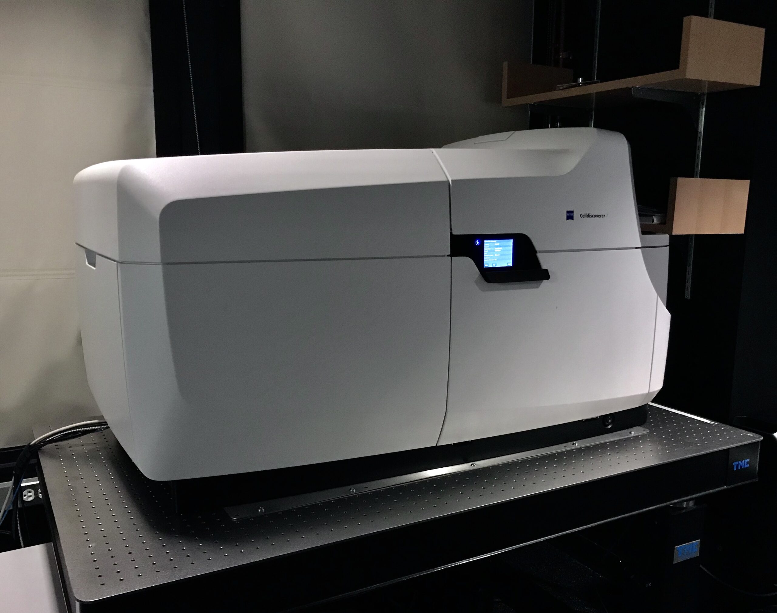
Zeiss Celldiscoverer 7 with LSM 900 (confocal) and Airyscan 2 (super-resolution)
Automated imaging platform. Especially suited to multi-well plate imaging in confocal, epifluorescence, and brightfield. Long-term, stable, time-lapse imaging capability of living cells and tissues. Accommodates glass slides, multi-chambered slides (up to 3 slides at a time), and Petri dishes of various sizes. Can perform high speed widefield epifluorescence in 3D with deconvolution. Airyscan 2 permits rapid mild super-resolution imaging of reliable 120 nm resolution using any fluorophores compatible with the lasers/filter sets.
Celldiscoverer 7 product information (Zeiss) [pdf]
Celldiscoverer7 training video (Youtube) [3 hr training session by Sebastian Ortiz (Zeiss) hosted by UC Berkeley CRL-MIC]
Automated features
- recognition and loading of sample container
- measurement of bottom thickness, material and skirt height
- UV disinfection of sample chamber
- adaptive correction of objective based on imaging depth
- autofocus and focus maps (hardware-based)
- active focus stabilization (hardware-based)
- barcode reading
- auto-immersion of objectives
- automated objective heating
- auto-adjustment of transmitted light contrast based on vessel geometry
- well plate calibration and adaptation of travelling ranges across sample chamber
- excitation adapts to FOV
Objectives
- Plan-Apochromat 5×/0.35 — WD 5.10 mm — thin vessel bottom 0.13-0.21 mm — thick vessel bottom up to 1.2 mm — 3 magnification changer options (2.5×/0.12) (5×/0.25) (10×/0.35)
- Plan-Apochromat 20×/0.7 [autocorr] — WD 2.20 mm — thin vessel bottom 0.13-0.21 mm — thick vessel bottom up to 1.2 mm — 3 magnification changer options (10×/0.35) (20×/0.7) (40×/0.7)
- Plan-Apochromat 20×/0.95 [autocorr] — WD 0.76 mm — thin vessel bottom 0.13-0.21 mm — 3 magnification changer options (10×/0.5) (20×/0.8) (40×/0.95)
- Plan-Apochromat 50×/1.2 [W] [autocorr] [autoimmersion] — WD 0.84 mm — thin vessel bottom 0.13-0.21 mm — 3 magnification changer options (25×/1.2) (50×/1.2) (100×/1.2)
Configuration highlights
- Afocal magnification changer (0.5× / 1× / 2×)
- Live cell incubation (temperature and CO2 control)
- Mounting frames for petri dishes, chamber slides, glass slides, multichamber and multiwell plates
- 7 LED excitation wavelengths for widefield imaging (385 nm, 420 nm, 470 nm, 520 nm, 567 nm, 590 nm, 625 nm)
- Multiple emission filter sets for widefield imaging (90 HE, 91 HE, 92 HE)
- Laser module URGB with 4 excitation wavelengths for confocal imaging (405 nm diode laser 5 mW | 488 nm diode laser 10 mW | 561 nm diode (SHG) laser 10 mW | 640 nm diode laser 5 mW)
- Transmitted light IR-LED (725 nm) brightfield and adaptive Phase Gradient Contrast
- Camera Axiocam 712 mono for widefield imaging (SONY IMX304 Exmor CMOS)
- LSM 900 scan-head with 2x GaAsP detectors and flexible spectral detection for confocal imaging
- AiryScan 2 detector (type 63×) for super-resolution imaging
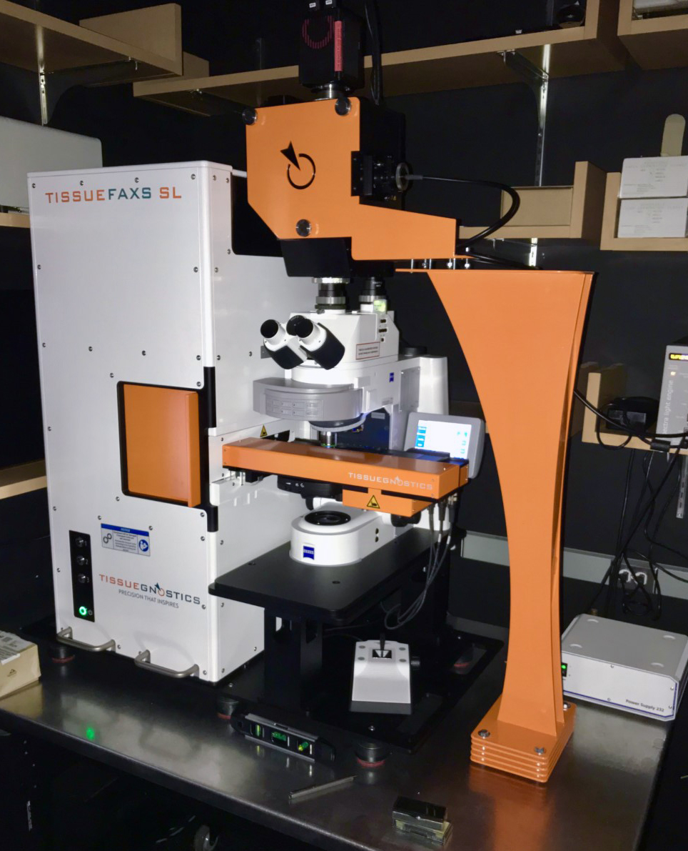
TissueGnostics TissueFAXS SL Q high-throughput slide-scanning confocal
Automated high-throughput confocal slide scanning system. Scans entire tissue sections or cultured cell populations on defined regions of slides at 5x, 10x, 20x, 40x, or 63x magnification in color brightfield, widefield fluorescence, and 3D confocal fluorescence. 5-color fluorescence imaging. Cell populations can be rapidly quantified using TissueQuest (fluorescence), HistoQuest (color brightfield), or StrataQuest (both, plus tissue structure quantification) analysis software. SL = 120-slide high-throughput loader. Q = spinning disc confocal. Instrument uses Zeiss AxioImager Z2 microscope stand.
TissueFAXS SL Q [tissuegnostics.com]
TissueFAXS platform [pdf]
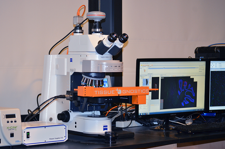
TissueGnostics TissueFAXS PLUS whole-slide scanner
Automated slide scanning system. Scans entire tissue sections or cultured cell populations on defined regions of slides at 5x, 10x, 20x, 40x, or 100x magnification in color brightfield and widefield fluorescence. 10 filter channel positions. Cell populations can be rapidly quantified with an approach similar to flow cytometry using TissueQuest (fluorescence), HistoQuest (color brightfield), or StrataQuest (both, plus tissue structure quantification).
TissueFAXS PLUS [tissuegnostics.com]
TissueFAXS platform [pdf]
Objectives
- Zeiss 2.5x EC Plan-NEOFLUAR 0.3NA air (for preview scanning)
- Zeiss 5x EC Plan-NEOFLUAR 0.16NA air
- Zeiss 10x EC Plan-NEOFLUAR 0.3NA air
- Zeiss 20x EC Plan-NEOFLUAR 0.5NA air
- Zeiss 40x EC Plan-Neofluar 0.75NA air
- Zeiss 100x EC Plan-Neofluar 1.30NA oil immersion
Filters (Filter cube position, suggested fluorophore, SpectraX excitation range, and Chroma cube specs)
- Position 1 (default) Transmitted light position
- Position 2 BV785 (395/25) ET395/25x T510lpxrxt ET785/60m
- Position 3 AF488 (470/24) ET470/30x T495lpxr ET515/30m
- Position 4 AF555 (550/15) ET550/15x T565lpxr ET625/30m
- Position 5 AF647 (640/30) ET640/30x T660lpxr ET705/
- Position 6 AF750 (740/20) ET740/40x T770lpxr ET780lp
- Position 7 BV421 (395/25) ET395/25x T400lp ET431/28m
- Position 8 BV570 (395/25) ET395/25x T510lpxrxt ET580/40m
- Position 9 BV650 (395/25) ET395/25x T655/40m T510lpxrxt
- Position 10 Multiband dichroic DAPI/FITC/TRITC/Cy5 (395/25, 470/24, 550/15, 640/30)
Cameras
- Baumer HXG40c (HX series) CMOS camera 16 bit (2048 × 2048) for brightfield imaging
- Hamamatsu Orca Flash 4.0 V2 cooled digital CMOS camera C11440-22CU for fluorescence imaging
Light source: Lumencor SPECTRA 3 Light Engine with eight solid state light sources each at 500mW per LED channel of the following excitation wavelength ranges:
- Violet – 390/22
- Blue – 440/20 (don’t have)
- Cyan – 475/34
- Teal – 510/25
- Green – 555/28
- Yellow – 585/35 (this is the Green LED position which can be interchanged with a 575/25 filter, so it won’t normally be used nor is it programmed into the TissueFAXS software)
- Red – 631/28
- NIR – 680/10
- Cy7 – 747/11 (factory-added option added in place of the 440/20)
Stage: Maerzhaeuser motorized scanning stage with 8-slide holders (for 1.0 × 3.0” and 1.5 × 3.0” glass slides)
Software:
- TissueFAXS image acquisition software.
- TissueQuest and HistoQuest image analysis software.
- StrataQuest advanced image analysis software.
Leica Stellaris confocal (non-core, coming soon to the core facility)
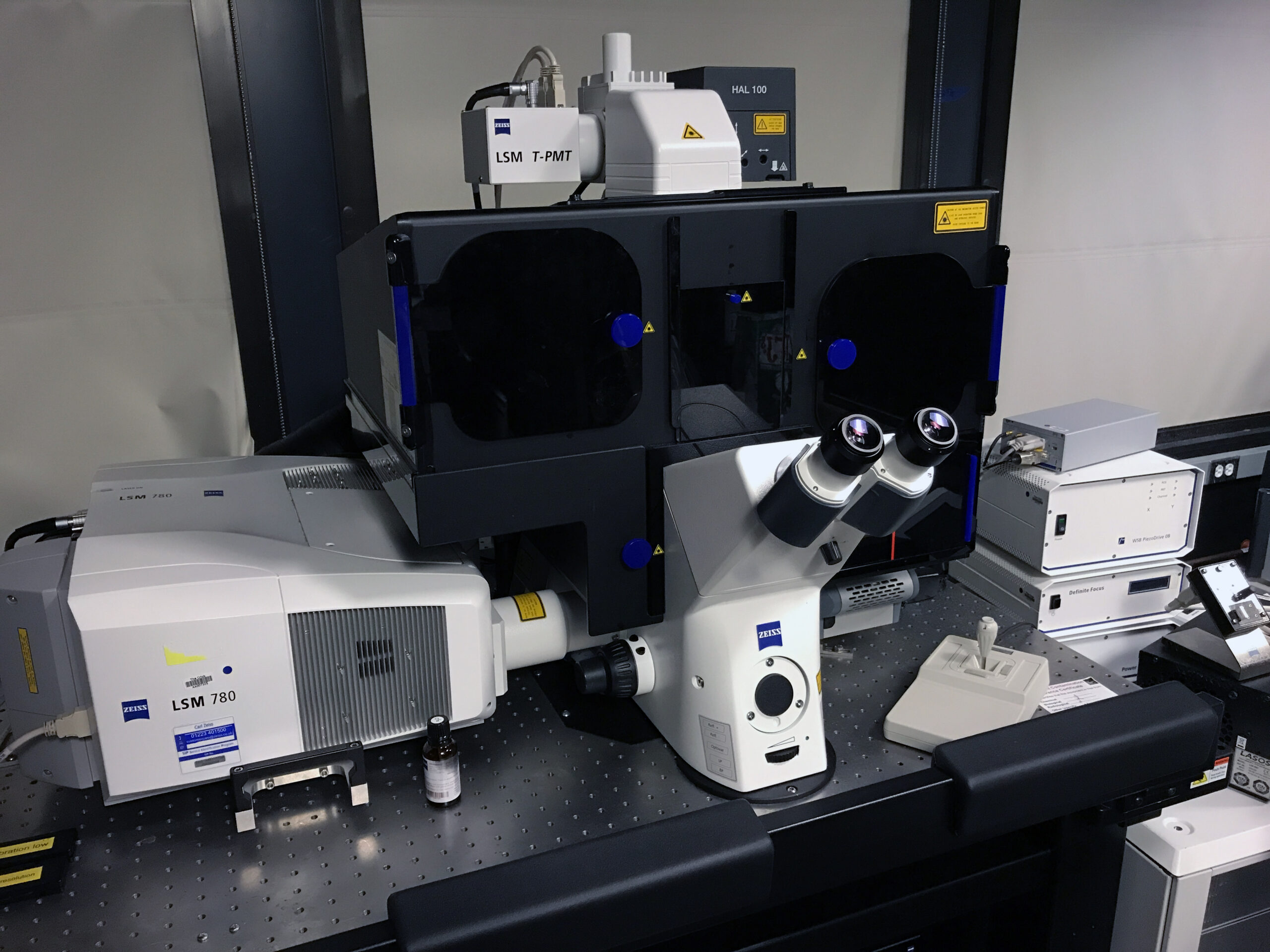
Zeiss Elyra PS.1 structured-illumination super-resolution (non-core, Ragon internal use only)
Confocal and structured-illumination super-resolution microscope (Zeiss Axio Observer 7 inverted, LSM780). Super-resolution capability includes structured illumination (SIM), photoactivated localization microscopy (PALM), 3D-PALM, total internal reflection (TIRF), and D-STORM compatibility. Up to eight-channel sequential acquisition. Up to four-channel simultaneous acquisition. For fixed and live cell imaging.
Zeiss Elyra PS.1 Brochure [PDF]
Zeiss Elyra PS.1 Sample prep guide [PDF]
Objectives
- 10x Zeiss Plan-APOCHROMAT air, 0.45NA
- 20X Zeiss Plan-APOCHROMAT air, 0.8NA
- 63X Zeiss Plan-APOCHROMAT oil, 1.4NA
- 63X Zeiss C APOCHROMAT water, 1.2NA
- 100X Zeiss Plan-APOCHROMAT oil, 1.46NA
- 40x Zeiss Plan-APOCHROMAT oil, 1.3NA
Laser Lines (Excitation Wavelengths)
- 405 nm: Diode laser, 50 mW
- 458, 477, 488 and 514 nm: Argon-ion laser, 30 mW
- 561 nm: Diode laser, 1 mW
- 633 nm: Helium-Neon laser, 15 mW
Primary Dichroic Filters
- NT 80/20
- HFT 405/488/561/633/KP 725
- HFT 405/488/561
- HFT 405/514/633
- HFT 458/514/561
- HFT 458/561
- HFT 458
- HFT 488
Secondary Dichroic Filters
- NFT490, NFT515, NFT565, NFT600, KP560
- NFT490, NFT515, NFT565
Barrier (Emission) Filters
- Ch 1) LP420, LP505, LP575, LP650, BP575-615 IR, BP575-630 IR, BP600-650
- Ch 2) LP420, LP475, LP505, BP420-480, BP475-545 IR, BP530-600, BP565-595, BP575-615 IR
- Ch 3) LP505, LP530, LP575, BP505-530, BP500-545 IR, BP530-600, BP565-595, BP575-615 IR
- Ch 4) BP530-600, BP575-615 IR, BP575-630 IR, BP600-650, LP530, LP575, LP600, LP650
Environmental control
- Incubator XL.S1 LSM
- Zeiss TempModule S1
- Zeiss CO2 module S1
- Heating device humidity S1
Accessories
- Ludl motorized scanning stage 130×100 DC
- X-CITE 120XL metal halide lamp
Software
- Zeiss “Zen” LSM Imaging Software