12-channel, 2-camera, 4-laser imaging flow cytometer for quantifying fluorescence signal and localization at the single cell level for cells in suspension.
Super resolution capability includes structured illumination (SIM), photoactivated localization microscopy (PALM), 3D PALM, TIRF, as well as being compatible for D-STORM. Up to eight channel sequential acquisition, and up to four channel simultaneous acquisition. Allows fixed or live cell imaging.
3D rapid imaging system using laser-based sheets of light to scan entire tissue mounts (wholemount) or small, cleared or- ganisms (thickness < 1 μm to 5 mm), with a 4-axis sample posi- tioning system and Multiview acquisition around different axial rotations. It has a CFP/YFP laser option in addition to the clari- ty option (20x objective).
A fully automated epifluorescence and confocal imaging platform that can accommodate slide, Petri dishes, chambered slides, and multiwell plates. It allows for fixed cell, live cell, and tissue imaging (2D/3D/4D), with automated water immersion objectives, and its own image analysis pipeline for high-throughput screening.
An older dual two photon (Mai Tai lasers) imaging system set up for live animal intravital fluorescence imaging of tissue.
An automated and high-speed slide scanner (up to 8 slides per session, at 10x, 20x, 40x, and 100x) with brightfield, epifluorescence, and multispectral scanning modes, which can create montages of stitched fields of view from tissue sections as well as cell cultured on coverslips. Cell populations can be gated according to their fluorescent marker profiles, similar to flow cytometry.
A high-speed, automated slide scanner (up to 120 slides per session) with brightfield, epifluorescence, and confocal scanning modes. The scanner can create montages of stitched fields of view from tissue sections as well as cell cultured on coverslips in either 2D or 3D z-stacks. It can scan at 10x, 20x, 40x, and 63x (oil immersion) in DAPI/FITC/Rhod/Cy5/AF750 channels (5 channel capability.)
| Microscopy Services (per hour) Effective 03/01/2023 | Partners of CFAR | External Academic | External Industry |
|---|---|---|---|
| TissueFAXS Confocal | $23.00 | $33.12 | $60.34 |
| CellDiscoverer 7 | $23.00 | $33.12 | $60.34 |
| TissueFAXS scanner | $18.00 | $25.92 | $47.22 |
| Microscope/Assisted/Training | $104.00 | $149.76 | $272.84 |
| ImageStream | $99.00 | $142.76 | $272.84 |
| ImageStream Assisted/Training | $132.00 | $190.08 | $346.30 |
| Analysis Assisted/Training | $104.00 | $149.76 | $272.84 |
All users are sent a monthly invoice. Users within Partners affiliates provide a grant number for billing through internal fund transfers initiated by the Core 7 days after invoices are sent. Users from outside Partners are sent an invoice with remittance instructions for payment via PO or check.
The Ragon Institute Imaging core has an online electronic management tool. Users are required to sign up and provide funding information for the payment of usage fees in order to be able to request training or schedule time on equipment.
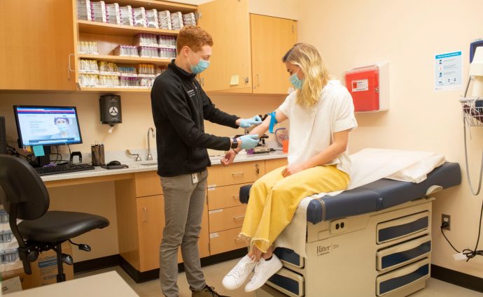
The Ragon’s clinical research focuses on clinical studies in infectious diseases such as HIV and COVID-19.
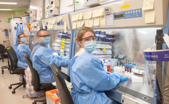
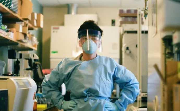
Our lab staff, comprised of students, postdocs, technicians, and scientists, is responsible for the hands-on research work at the Ragon.
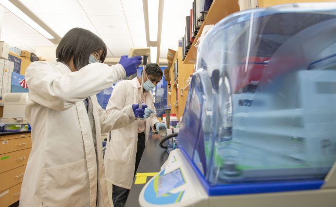
The Ragon’s research creates knowledge from our collaborative, cross-disciplinary approach, breaking down the silos of academia.